An elderly patient lies still inside the humming bore of an MRI machine, the images slowly rendering a detailed map of their brain. When the results return, the radiologist points to a tiny, fluid-filled sac in the center of the brain—a small cyst. The medical team dismisses it as an incidental finding, a common byproduct of aging that requires no intervention. Yet the patient, who has spent the last several months battling a precipitous decline in sleep quality and a persistent sense of nocturnal restlessness, wonders if this small void is actually the epicenter of their insomnia.
The Biological Blueprint of Pineal Degeneration
The pineal gland serves as the body's primary endocrine clock, responsible for the synthesis and secretion of melatonin, the hormone that regulates circadian rhythms. Its cellular architecture is highly specialized and remarkably skewed. Approximately 95 percent of the gland consists of pinealocytes, the primary cells dedicated to melatonin production. The remaining 5 percent is composed of a supporting cast of astrocytes, which provide structural and nutritional stability, and microglia, which handle the immune response within the brain. These cells are embedded within a complex network of blood vessels and nerve fibers, creating a delicate biological machine.
Recent data published in a study [https://doi.org/10.3390/ijms27073093] reveals that this machine does not simply wear out; it structurally collapses. The research indicates that the lobular organization of the pineal gland undergoes a process of degradation as a human ages. This trajectory is not unique to the brain. It closely mirrors the involution seen in the thymus, where T-cells mature, and the structural breakdown observed in lymph nodes during the aging process. The study specifically categorizes the correlation between the collapse of these lobular structures and the behavior of the resident astrocyte populations, suggesting that the very scaffolding of the gland fails over time.
The Functional Erosion of the Third Eye
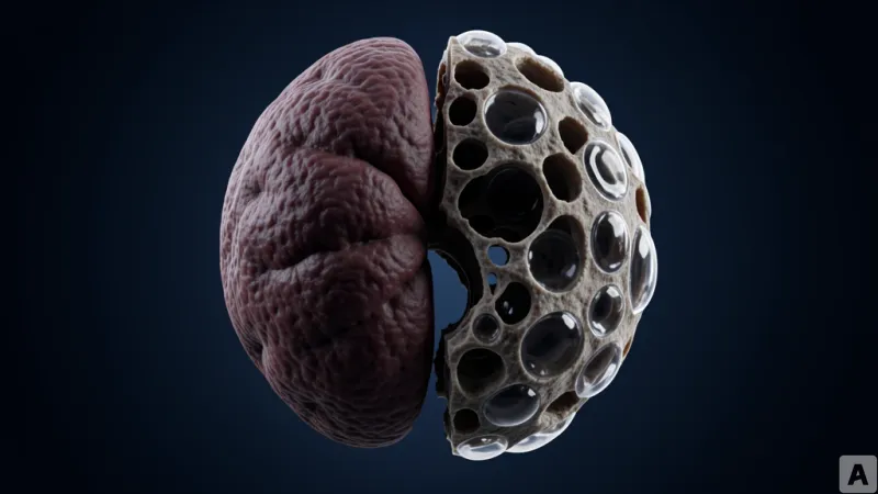
For decades, the pineal gland has been shrouded in mysticism, often referred to as the third eye or a gateway to higher consciousness. However, the biological data presents a far more clinical and cold reality: the gland is subject to inevitable physical decay. The most visible marker of this decline is the emergence of glial cysts—fluid-filled pockets originating from the supporting glial cells. While these cysts appear in patients across various age groups and are typically asymptomatic from a neurological perspective, their presence signals a deeper functional crisis.
The critical distinction lies in the loss of parenchyma, the actual functional tissue of the organ. The data suggests that glial cysts do not simply appear in empty space; they emerge when the lobular structure collapses and the astrocyte network becomes sparse. As the structural integrity of the gland vanishes, these cysts expand, effectively colonizing the space where melatonin-producing pinealocytes once resided. This is not a case of the cells simply stopping their work, but rather a physical displacement where the machinery of hormone production is crowded out by biological voids.
This mechanism explains why the decline in melatonin is often gradual yet relentless. While certain pathological conditions can accelerate this decay, the evidence indicates that normal aging is the primary driver. The loss of sleep quality in the elderly is not merely a psychological shift or a change in habit, but the direct result of a structural failure. The internal architecture of the pineal gland collapses under the weight of time, leading to a functional deficit that no amount of sleep hygiene can fully resolve.
The degeneration of the pineal gland is not a disease to be cured, but a biological clock ticking toward an inevitable structural collapse.