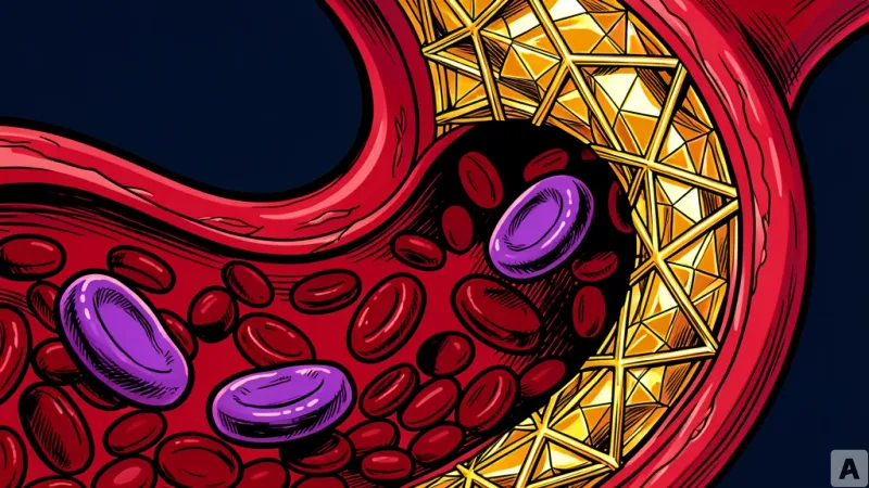
A cardiologist stares at two nearly identical CT scans on a high-resolution monitor. Both images show an abdominal aortic aneurysm, where the primary artery of the body has weakened and ballooned outward like a fragile bubble. To the naked eye, the risk profiles look the same. Yet, in the clinical reality of the ward, one of these patients will remain stable for a decade, while the other will suffer a catastrophic rupture within months. For years, the medical community has operated in this blind spot, relying on the diameter of the vessel as the sole metric for surgical intervention, effectively guessing which ticking time bomb will explode first.
The Genetic Trigger in the Blood
Recent analysis of 44 patients who underwent surgery for abdominal aortic aneurysms reveals that the secret to this volatility is not in the vessel wall itself, but in the blood. The research indicates that approximately 60% of these patients carry Clonal Hematopoiesis, a phenomenon where blood-forming stem cells accumulate mutations as part of the aging process. The data shows a direct correlation between the presence of these mutations and the acceleration of aneurysm growth, suggesting that the blood is actively signaling the vessel to degrade.
To uncover the mechanism, researchers utilized animal models featuring mutations in Tet2, a gene responsible for regulating the differentiation of blood cells. The findings reveal a disturbing cellular transformation. Macrophages, the immune cells typically tasked with clearing cellular debris, begin to exhibit characteristics of Osteoclasts, the specialized cells that break down bone tissue. This transformation triggers an overproduction of MMP9, a matrix metalloproteinase 9 that aggressively degrades the structural proteins of the vessel wall. As MMP9 levels rise, the aorta loses its elasticity and thins, transforming a stable bulge into a critical failure point.
Hijacking the Bone-Degradation Pathway
The discovery shifts the conversation from simple vascular wear-and-tear to a systemic biological hijacking. The process of vessel destruction is governed by the RANK/RANKL signaling pathway, a system evolved specifically to manage the balance of bone formation and resorption. In a healthy body, this pathway ensures skeletal integrity; however, in patients with these specific blood mutations, the body mistakenly deploys this bone-eating machinery within the aorta. The vessel is not simply wearing out; it is being actively digested by immune cells that believe they are breaking down bone.
This realization opens a door to drug repurposing, bypassing the decade-long timeline of traditional drug development. Because the RANK/RANKL pathway is already the primary target for osteoporosis treatments, researchers tested existing medications on mouse models. The results were definitive. The administration of Anti-RANKL antibodies and Alendronate, a bisphosphonate drug used to prevent bone loss, significantly inhibited the growth of the aneurysms. By blocking the signal that tells macrophages to act like bone-destroyers, the drugs stabilized the vessel wall and halted the progression of the disease.
This approach transforms the clinical management of aortic aneurysms from a reactive game of measurement to a proactive biological strategy. By integrating blood-based mutation screening with existing FDA-approved therapies, clinicians can now identify high-risk patients long before their vessels reach a critical diameter. This removes the guesswork from the operating room, allowing doctors to avoid unnecessary early surgeries for stable patients while providing life-saving pharmacological intervention for those whose blood is programmed to destroy their arteries.
Medical science is now recognizing that the aging of the blood can trigger identical destructive mechanisms across entirely different organ systems.