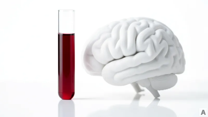
The annual physical is usually a ritual of mundane metrics: blood pressure, cholesterol, and a standard blood draw that most patients ignore until the results hit their inbox. For decades, these routine panels have been used to track heart health or metabolic dysfunction, while the brain remained a black box, largely inaccessible until cognitive decline became impossible to ignore. But a shift is occurring in the clinical landscape. The same blood sample used to check for a common infection may now hold the key to predicting a patient's cognitive fate years before the first sign of memory loss appears.
The 400,000-Patient Dataset and the NLR Marker
On April 3, a research team from NYU Langone Health published a landmark study in the journal Alzheimer's & Dementia, revealing a potent correlation between the Neutrophil-to-Lymphocyte Ratio (NLR) and the risk of developing dementia. The scale of the study provides a level of statistical power rarely seen in early-detection research. The team analyzed data from nearly 400,000 individuals, comprising 285,000 patients across four NYU Langone hospitals and an additional 85,000 patients from the Veteran's Health Administration. The cohort focused specifically on adults aged 55 and older who had not yet been diagnosed with Alzheimer's or any other form of dementia at the time of their initial NLR measurement.
NLR is not a specialized or expensive test; it is derived from a Complete Blood Count (CBC), one of the most common laboratory tests in modern medicine. The ratio is calculated by dividing the number of neutrophils—white blood cells that act as the body's first responders to infection and inflammation—by the number of lymphocytes, which are responsible for regulating the immune response and maintaining immunological memory. The findings were stark: patients with NLR levels above the median showed a consistently higher probability of developing dementia, including Alzheimer's disease.
Interestingly, the correlation was not uniform across all demographics. The research highlighted a stronger link between elevated NLR and dementia risk among women and patients of Hispanic descent. This suggests that systemic inflammation may interact with genetic or environmental factors in ways that disproportionately affect certain populations. The study was supported by several National Institutes of Health (NIH) grants, including R01AG092953 and R01AG070821, alongside funding from the Alzheimer's Association and the BrightFocus Foundation. To move beyond correlation, the researchers are now utilizing the VIDA lab (Vascular and Immune Dysfunction lab) to integrate PET scans, which visualize brain metabolic activity, with diffusion MRI, which analyzes the microstructure of brain tissue by tracking water molecule movement. This combined approach aims to determine if neutrophils are actively driving the degradation of cognitive function.
The Biological Tension Between Protection and Destruction
To understand why a ratio of white blood cells in the arm can predict the death of neurons in the brain, one must look at the immune system as a balancing act. In a healthy state, neutrophils and lymphocytes work in tandem to protect the body. Neutrophils are the infantry; they arrive quickly at the site of an injury or infection to neutralize threats. Lymphocytes are the strategists; they coordinate the long-term defense and ensure the system returns to homeostasis once the threat is gone.
When the NLR rises, it signals a state of systemic imbalance. Using a fire-fighting analogy, neutrophils are like fire trucks. In a controlled emergency, they are essential for putting out the fire and preventing the spread of damage. However, if fire trucks are deployed in massive numbers to areas where there is no active fire, or if they refuse to leave after the fire is out, they begin to block traffic, crush sidewalks, and damage the very infrastructure they were meant to protect. In the human body, overactive neutrophils trigger chronic inflammation. When this inflammation persists, it can breach the blood-brain barrier and damage the delicate neural architecture of the brain.
Evidence from Alzheimer's patients already shows signs of neutrophil-driven inflammation within brain tissue. Animal models have further confirmed that these cells can accelerate the progression of the disease. The danger is compounded by the aging process; as humans age, the body's ability to clear out old, dysfunctional neutrophils diminishes, leaving the brain vulnerable to a slow-motion inflammatory assault.
This discovery represents a fundamental reversal in the diagnostic timeline of dementia. Historically, the medical community has been reactive, initiating diagnostic protocols only after a patient exhibits symptomatic cognitive decline—at which point significant neuronal loss has already occurred. The NLR marker, however, spikes long before the patient forgets a name or loses their way home. While a high NLR is not a definitive diagnosis of Alzheimer's on its own, it functions as a critical gateway tool. By combining this low-cost blood marker with other risk factors, clinicians can identify high-risk individuals who require immediate, high-resolution screening via PET or MRI, effectively moving the window of intervention forward by several years.
There are still technical hurdles to clear. Neutrophils have an incredibly short lifespan, meaning researchers must work with fresh blood samples to maintain accuracy. The current scientific frontier is determining whether the NLR is merely a biomarker—a smoke detector warning of a fire—or a causal agent—the match that starts the fire. If neutrophils are proven to be a direct cause of cognitive decline, the focus of Alzheimer's treatment could shift from clearing amyloid plaques to modulating the activity of these specific immune cells.
The transition from expensive, invasive brain imaging to a simple blood ratio transforms the immune system into the most accessible window we have into the future of brain health.