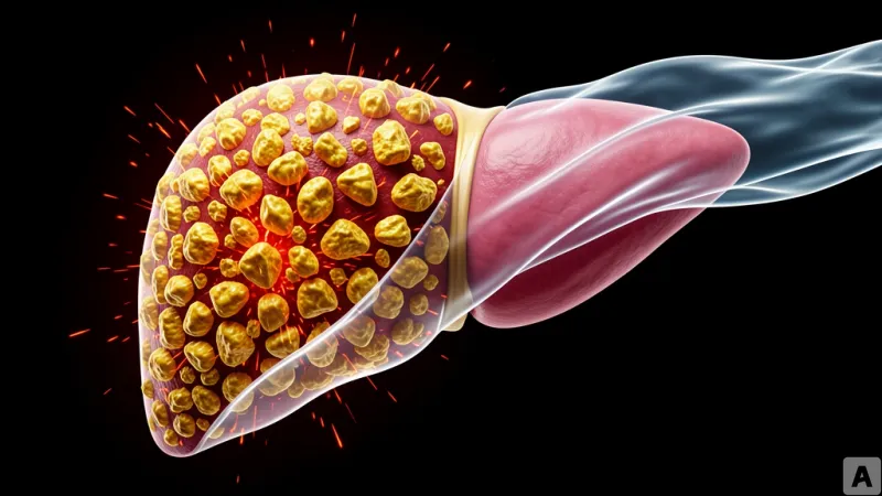
A clinical biopsy report arrives with a frustratingly common pattern. The patient shows high levels of chronic inflammation in the liver tissue, yet there is no clear primary cause. To a clinician, it looks like the inevitable toll of aging, but the degree of hepatocyte degeneration is too severe to be dismissed as simple senescence. Standard anti-inflammatory treatments are administered, but the response is sluggish, leaving the underlying pathology to simmer. This gap between observed inflammation and therapeutic failure suggests that the medical community is missing a specific cellular culprit.
The Molecular Blueprint of Macrophage Senescence
To isolate the cause of this persistent inflammation, researchers focused on Bone Marrow-Derived Macrophages (BMDMs), inducing cellular senescence through two distinct stressors: Ionizing Radiation (IR) and the chemotherapy agent Doxorubicin (Doxo). The results revealed a precise molecular shift. Ten days after treatment, the cells showed a marked decrease in the expression of Ki-67, a primary marker for cell proliferation, and Lamin B1, a critical protein that maintains the structural integrity of the nuclear envelope. Simultaneously, the levels of pNF-κB, a protein that regulates inflammatory responses, increased, and the activity of SA-β-gal, the gold-standard marker for senescent cells, spiked.
The physical transformation of these cells was equally stark. The macrophages entered a state of cell cycle arrest at the G2-M phase, resulting in a polyploid state where cells possessed a genome of 4N or greater. When visualized using DAPI imaging, the control macrophages maintained uniform nuclear size and shape. In contrast, the IR and Doxo-treated groups exhibited enlarged, irregularly shaped nuclei. The timeline of this arrest was captured using CellTrace Violet, a dye used to track cell division. On the first day following IR treatment, approximately 20% of the cells continued to divide. However, between day five and day ten, all cellular division ceased entirely.
These senescent macrophages were defined by a unique expression profile: the simultaneous presence of p21, a cell cycle inhibitor, and TREM2 (Triggering Receptor Expressed on Myeloid cells 2), an immune receptor. The research identified two primary catalysts for this transition: accumulated DNA damage and the excessive accumulation of cholesterol. This specific p21+ TREM2+ phenotype was not limited to lab-grown cells. The team found these macrophages concentrated in the livers of aged mice, mice in MASLD (Metabolic dysfunction-associated steatotic liver disease) models, and within human liver cirrhosis tissues.
Beyond M1 and M2: The Irreversible Inflammatory Driver
For decades, macrophage research has been dominated by the paradigm of polarization, categorizing cells as either M1 (pro-inflammatory) or M2 (anti-inflammatory). This framework assumes a level of plasticity, where a cell can shift its state based on the environment. The discovery of p21+ TREM2+ macrophages shatters this binary by introducing a state of irreversible senescence. Unlike M1 or M2 polarization, which are reversible functional states, senescence is a permanent exit from the cell cycle that transforms the cell into a chronic source of toxicity.
There is a biological irony in why macrophages are so susceptible to this state. Macrophages are naturally resilient to radiation and possess high metabolic flexibility, allowing them to survive in extreme stress environments where other cells would perish. However, this very resilience becomes a liability. Because they survive the initial insult, they accumulate massive amounts of DNA damage and metabolic stress over time, eventually tipping into a senescent state that they cannot escape.
The mechanism driving this toxicity is the Senescence-Associated Secretory Phenotype (SASP). In these p21+ TREM2+ cells, the SASP is not a simple immune response but is driven by the leakage of mitochondrial DNA into the cytoplasm. This triggers Type I interferon signaling, creating a feedback loop of persistent inflammation. The cell is no longer acting as a defender of the tissue but as a permanent beacon of inflammatory signals that damage surrounding healthy hepatocytes.
When the researchers applied Senolytic therapy—drugs designed to selectively eliminate senescent cells—the results were immediate. By removing the p21+ TREM2+ population, both aged mice and MASLD model mice showed a significant reduction in liver inflammation and steatosis. This proves that these specific macrophages are not merely bystanders of liver aging but are the primary engines driving the disease. The pathology is not caused by a lack of healthy immune function, but by the active, malicious presence of these irreversible senescent cells.
Immune senescence must be redefined as an active driver of disease rather than a passive decline in function.